Friday 18 November 2011

Photomicrography


Nikon Small World 
Photomicrography Competition

The Nikon Small World Photomicrography Competition lets us see beyond the capabilities of our unaided eyes. Almost 2000 entries from 70 countries vied for recognition in the 37th annual contest, which celebrates photography through a microscope. Images two through 21 showcase the contest's winners in order, and are followed by a selection of other outstanding works. Scientists and photographers turned their attention on a wide range of subjects, both living and man-made, from lacewing larva to charged couple devices, sometimes magnifying them over 2000 times their original size. -- Lane Turner
Dr. Igor Siwanowicz of the Max Planck Institute of Neurobiology in Martinsried, Germany shot "Portrait of a Chrysopa sp. (green lacewing) larva" at 20x magnification using the confocal method. (Dr. Igor Siwanowicz)
A marine copepod, Temora longicornis, at 10x magnification imaged by Dr. Jan Michels of Christian-Albrechts-Universität zu Kiel in Kiel, Germany using the confocal process with autofluorescence and Congo red fluorescence. (Dr. Jan Michels)

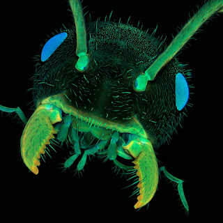
An ant head magnified ten times and photographed by Dr. Jan Michels of Christian-Albrechts-Universität zu Kiel in Kiel, Germany using the confocal method with autofluorescence. (Dr. Jan Michels)

Using reflected light, Yanping Wang of the Beijing Planetarium in Beijing captured sand at 4x magnification. (Yanping Wang)

A freshwater shrimp eye and head shot with image stacking photography by Jose R. Almodovar at the Microscopy Center, Biology Department, UPR Mayaguez Campus in Mayaquez, Puerto Rico. (Jose R. Almodovar)

Bladderwort bladder (Utricularia gibber) photographed with the darkfield method and magnified 40x by Jose R. Almodovar at the Microscopy Center, Biology Department, UPR Mayaguez Campus in Mayaquez, Puerto Rico. (Jose R. Almodovar)

A confocal image of giant liposomes of pulmonary surfactant was magnified 40 times by Dr. Jorge Bernardino de la Serna of MEMPHYS - Center for Biomembrane Physics, Department of Biochemistry and Molecular Biology in Odense, Denmark. (Dr. Jorge Bernardino de la Serna)

Confocal image of Clausidium sp. nov., female with egg sacs, ventral view was made by Dr. Terue Kihara of the German Centre for Marine Biodiversity Research (DZMB) in Senckenberg am Meer, Germany. (Dr. Terue Kihara)

An antique mount of a blowfly (Calliphoridae) proboscis was photographed by Dr. Davis Linstead of Kent, UK with differential interference contrast. (Dr. David Linstead)

The double compound eyes of a male St. Mark's fly (Bibio marci) photographed with reflected (episcopic) diffuse illumination by Dr. David Maitland of Feltwell, UK. (Dr. David Maitland)

No comments:

Post a Comment

Silahkan komentar dengan santun & tidak mengandung spam, terima kasih.